
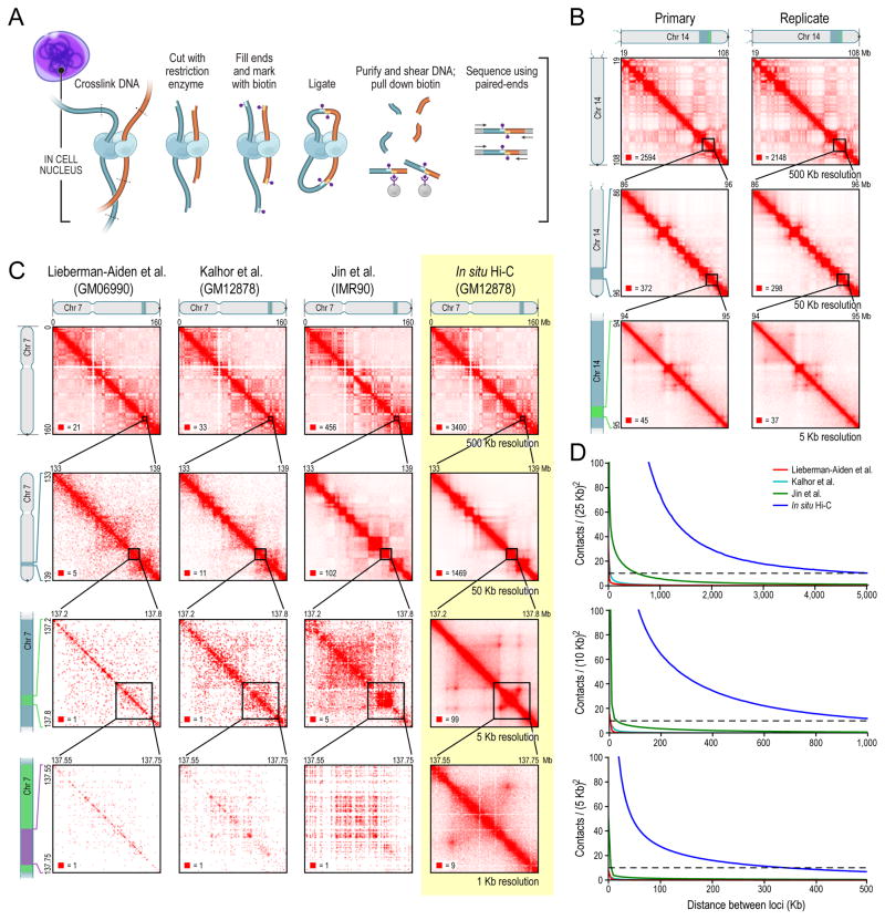
We use in situ Hi-C to probe the 3D structure of genomes, developing haploid and diploid maps of 9 cell sorts. The densest, in human lymphoblastoid cells, incorporates 4.9 billion contacts, reaching 1 kb resolution. We discover that genomes are partitioned into contact domains (median size, 185 kb), that are related to distinct patterns of histone marks and segregate into six subcompartments. We determine ∼10,000 loops.
These loops often hyperlink promoters and enhancers, correlate with gene activation, and present conservation throughout cell sorts and species. Loop anchors usually happen at area boundaries and bind CTCF. CTCF websites at loop anchors happen predominantly >>90%) in a convergent orientation, with the uneven motifs “going through” each other. The inactive X chromosome splits into two large domains and incorporates massive loops anchored at CTCF-binding repeats.
The 160-kilobase genome of the bacterial endosymbiont Carsonella.
Previous research have prompt that the minimal mobile genome could possibly be as small as 400 kilobases. Here, we report the full genome sequence of the psyllid symbiont Carsonella ruddii, which consists of a round chromosome of 159,662 base pairs, averaging 16.5% GC content material. It is by far the smallest and most AT-rich bacterial genome but characterised.
The genome has a excessive coding density (97%) with many overlapping genes and decreased gene size. Genes for translation and amino acid biosynthesis are comparatively effectively represented, however quite a few genes thought of important for all times are lacking, suggesting that Carsonella could have achieved organelle-like standing.
Stable transfection of the human parasite Leishmania main delineates a 30-kilobase area ample for extrachromosomal replication and expression.
To delineate segments of the genome of the human protozoan parasite Leishmania main essential for replication and expression, we developed a vector (pR-NEO) which could be reproducibly launched into L. main. This DNA was derived from a 30-kilobase extrachromosomal amplified DNA bearing the dihydrofolate reductase-thymidylate synthase gene, with the coding area for neomycin phosphotransferase substituted for that of dihydrofolate reductase-thymidylate synthase and a bacterial origin of replication and selectable marker added. G418-resistant traces have been obtained at excessive effectivity by electroporation of pR-NEO (approaching 10(-4) per cell), whereas constructs bearing an inverted neo gene or missing Leishmania sequences didn’t confer resistance. pR-NEO replicated in L. main and gave rise to accurately processed transcripts bearing the trans-spliced miniexon. Molecular karyotype evaluation confirmed that in some traces pR-NEO DNA exists completely as an extrachromosomal circle, a discovering supported by the rescue of intact pR-NEO after transformation of Escherichia coli.
These information genetically localize all parts required in cis for DNA replication, transcription, and trans splicing to the Leishmania DNA contained inside pR-NEO DNA and sign the introduction of secure transfection methodology for addressing molecular phenomena in trypanosomatid parasites.